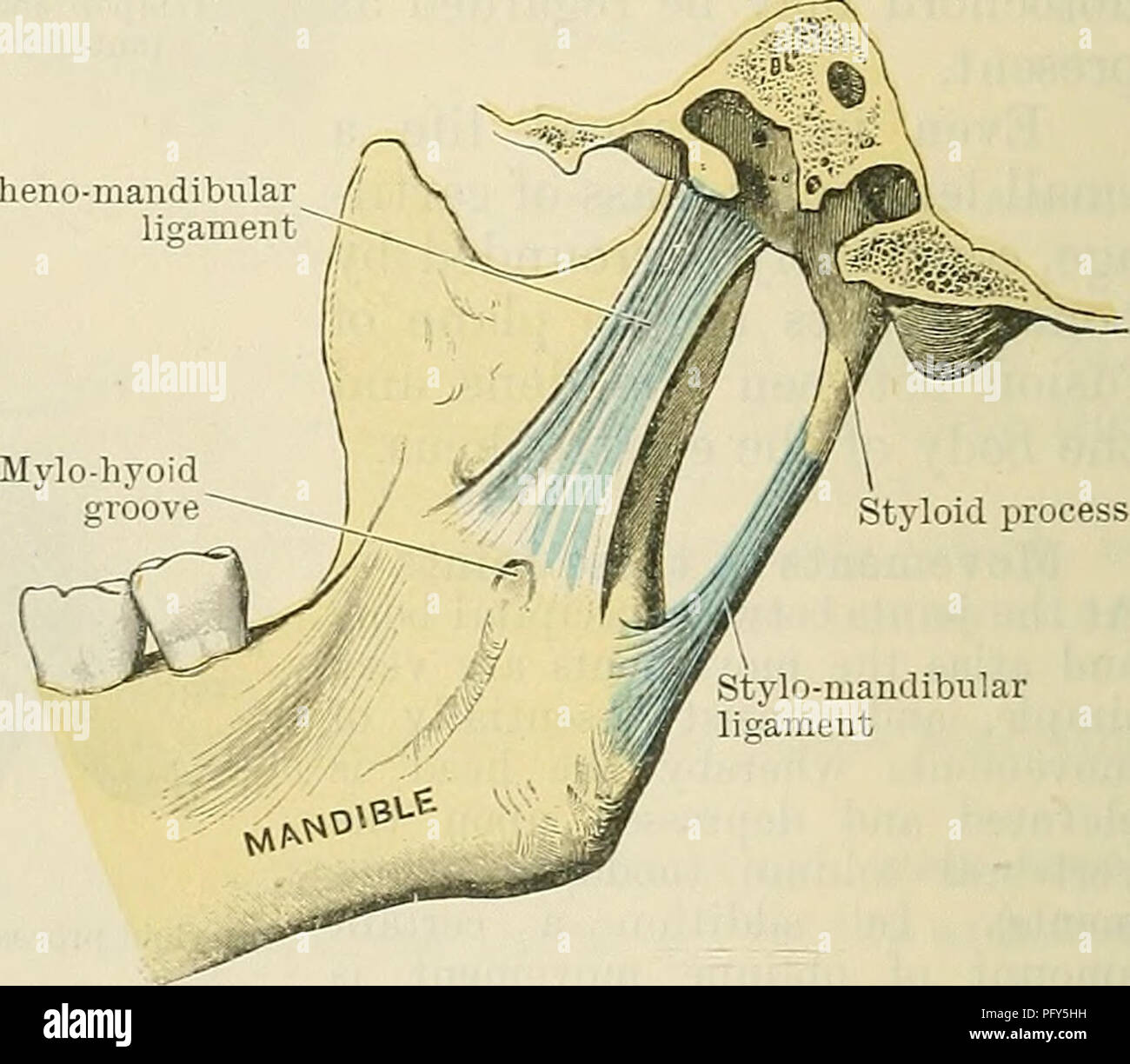
. Cunningham's Text-book de l'anatomie. L'anatomie. Fig. 299. de la mandibulaire. L'O.T. mandibulaire (mâchoire) est un arthrodial diarthrosis. Il se produit entre la fosse mandibulaire de l'os temporal et du condyle de la mandibule. Ces deux surfaces articulaires sont nettement dissemblables tant dans la taille et la forme. Dans ses grandes lignes la surface articulaire de la tête de la mandibule est cylindrique, ayant l'axe dirigé du côté médial et latéral vers l'avant. D'autre part, la fosse mandibulaire est concavo-convexes de l'arrière vers l'avant. Son su articulaire
1692 x 1476 px | 28,7 x 25 cm | 11,3 x 9,8 inches | 150dpi
Informations supplémentaires:
Cette image appartient au domaine public, ce qui signifie que le droit d’auteur a expiré ou que le titulaire du droit d’auteur a renoncé à ses droits. Les frais facturés par Alamy couvrent l’accès à la copie haute résolution de l’image.
Cette image peut avoir des imperfections car il s’agit d’une image historique ou de reportage.
. Cunningham's Text-book of anatomy. Anatomy. Fig. 299.—Section through the Mandibular Joint. The mandibular joint (O.T. temporomandibular) is an arthrodial diarthrosis. It occurs between the mandibular fossa of the temporal bone and the condyle of the mandible. These two articular surfaces are markedly dissimilar both in size and shape. In its general outline the articular surface of the head of the mandible is cylindrical, having its long axis directed from the medial side laterally and forwards. On the other hand, the mandibular fossa is concavo-convex from behind forwards. Its articular surface includes the tuberculum articulare—the eminence at the base of the anterior root of the zygoma. The articular surfaces of the bones are clothed with hyaline en- crusting cartilage, whilst the articular cavity is divided into a superior and inferior part by a disc of fibro-cartilage. Ligaments.—The joint is invested by an articular capsule which is quite com- plete, but is very thin on the medial side. The lateral part of the fibrous stratum of the capsule—the temporo-mandibular liga- ment (O.T. external lateral) (Fig. 298)—is divisible into anterior and posterior portions which are attached superiorly to the root tubercle and inferior border of the zygomatic process of the temporal bone, and inferiorly to the lateral side and posterior border of the neck of the mandible. The direction of its fibres is downwards and backwards. Within the capsule there is a disc of fibro-cartilage, the discus articularis (Fig. 299), which is moulded upon the condyle of the mandible below, and on the articular surface of the temporal bone above. It thus compensates for the incongruity between the articular surfaces of the two bones. The disc is attached circumferentially to the capsule. It is widest in the trans- verse direction, thicker posteriorly than anteriorly, and thinnest towards the centre, where it may be perforated. Its anterior margin is intimately associated with the in